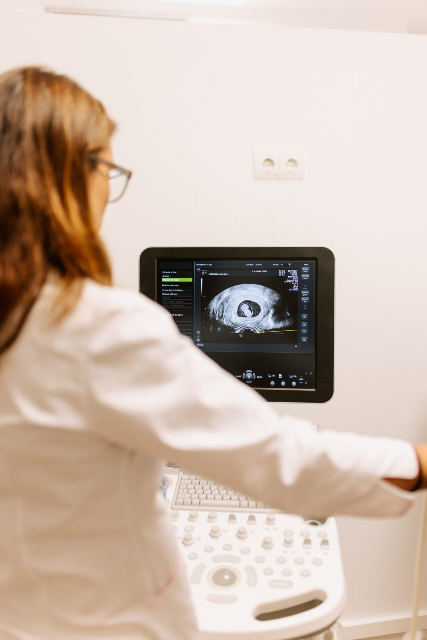
Gynecological ultrasounds
From cancer prevention to prenatal diagnosis, ultrasound is a safe and painless way to view the status of internal organs.
Ultrasounds
In these, the structures of the body are represented as images thanks to ultrasound waves. Ultrasound is the technique par excellence for diagnosing any gynecological pathology. The dynamic and direct exploration advantages provide greater reliability than other complementary diagnostic techniques such as CT or magnetic resonance imaging.


Gynecological vaginal and abdominal ultrasounds are a tool that allows us to obtain a detailed visualization of the internal genital organs for later evaluation. These ultrasounds are essential for detecting and monitoring a wide variety of gynecological conditions, including fibroids, ovarian cysts, polycystic ovaries, ovarian cancer and endometrial problems.
2D obstetric ultrasound
This is the type of ultrasound most commonly used to monitor growth and detect possible fetal abnormalities. At 20 weeks of pregnancy, when the fetus is fully formed, a morphological ultrasound is performed to determine that it does not suffer from any malformations. During the gestation period, it is common to perform multiple ultrasounds periodically, including a Doppler ultrasound, which makes it possible to monitor and observe the blood flows that feed the fetus.


4D obstetric ultrasound
4D ultrasounds allow you to see the surface of the fetus thanks to a computerized process of ultrasound signals. Seeing the surface means being able to appreciate the physiognomy of the fetus's face, a very gratifying fact for those expecting a child. They are known as 4D because movement is introduced and considered as a fourth dimension.
Gynecological Ultrasounds
Gynecologic ultrasounds are essential for the evaluation and management of various gynecologic conditions. This noninvasive procedure allows physicians to obtain detailed images of the uterus, ovaries, and endometrium, facilitating the detection of:
- Uterine fibroids: Benign tumors in the wall of the uterus, the size, location and number of which can be accurately assessed.
- Ovarian cysts: Fluid-filled sacs in the ovaries, whose nature (functional or pathological), size and evolution can be monitored.
- Polycystic Ovary Syndrome (PCOS): Condition in which the ovaries contain multiple small cysts, identifiable and monitorable through ultrasound.
- Ovarian Cancer: Identification of suspicious ovarian masses that require further evaluation.
- Endometrium: Evaluation of the thickness and structure of the uterine lining to diagnose conditions such as endometrial hyperplasia and endometrial cancer.

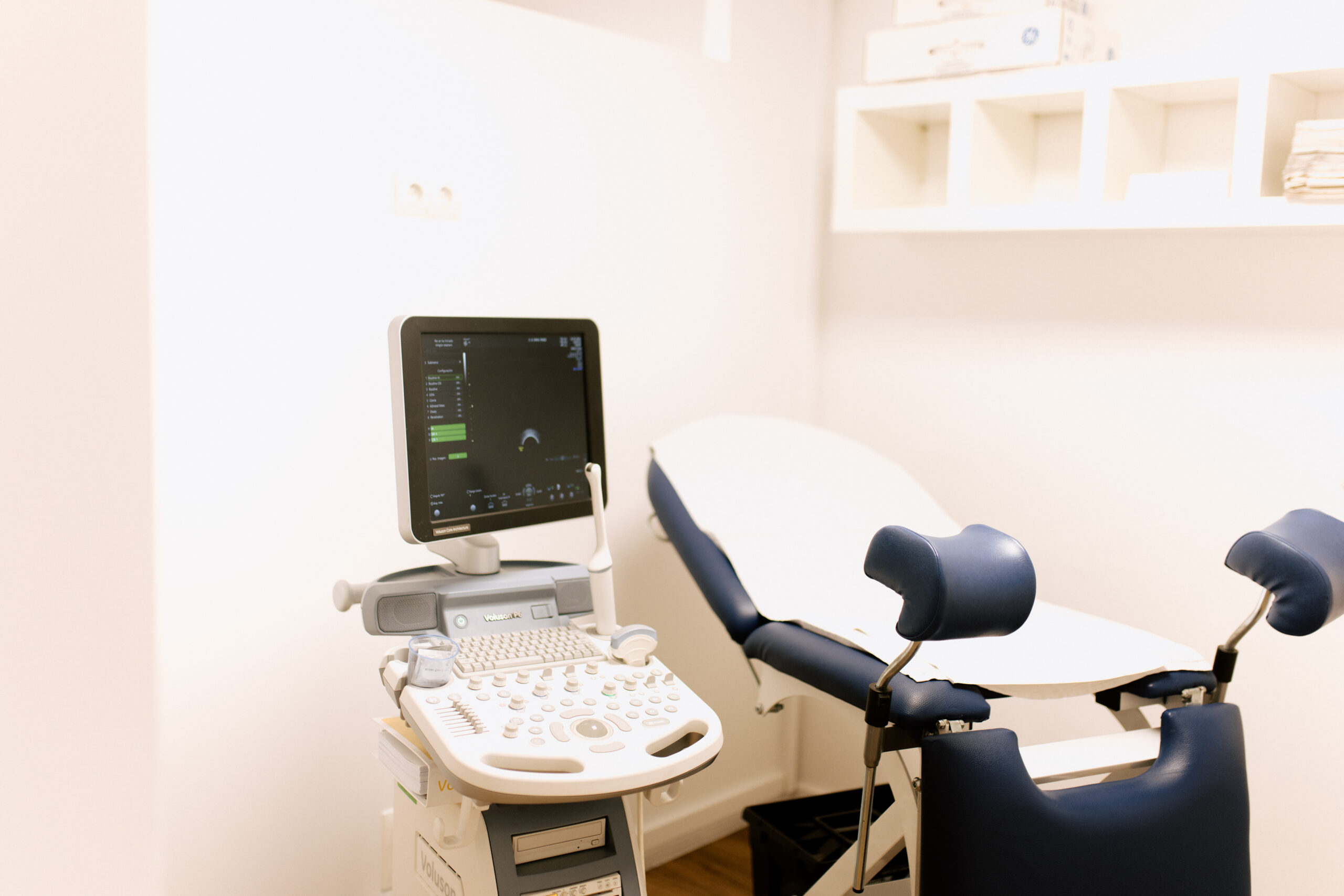
Gynecological Ultrasound Procedure
The gynecologic ultrasound procedure is simple and usually noninvasive. It can be performed transabdominally, through the abdominal wall, or transvaginally, using a special device inserted into the vagina. Depending on the type of ultrasound, specific instructions may be given, such as drinking water before the exam to fill the bladder in the case of a transabdominal ultrasound.
The images obtained are interpreted to identify any abnormalities, and the doctor will discuss the results with you to plan the next steps based on the findings. Gynecological ultrasounds are an indispensable tool for the evaluation and management of various gynecological conditions, and at GINEPEREZ, we strive to offer the best care with advanced technologies and a personalized approach.